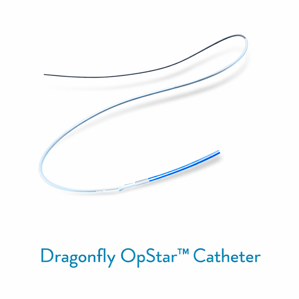
The Dragonfly™ OpStar Imaging Catheter is a high-performance optical coherence tomography (OCT) catheter designed for detailed intravascular imaging during coronary interventions.
Enables high-resolution, real-time visualization of coronary artery structures to support accurate assessment of vessel morphology, lesion characteristics, and stent placement.
Engineered for smooth deliverability and consistent imaging quality across a wide range of coronary anatomies.
Key Features
High-Resolution OCT Imaging: Provides superior visualization of vessel structures for precise assessment and procedural planning.
Smooth Deliverability: Designed to navigate tortuous coronary anatomy with ease and flexibility.
Optimized Optical and Mechanical Performance: Ensures consistent high-quality imaging across a wide range of coronary anatomies.
System Compatibility: Compatible with OPTIS™ and OPTIS™ Next OCT systems for seamless integration.
Single-Use, Sterile Design: Ensures safety and reliability during procedures.
Procedural Guidance: Provides detailed imaging to guide stent placement and post-deployment evaluation.
Clinical Applications
Detailed intravascular imaging of coronary arteries
Assessment of vessel morphology and lesion characteristics
Guiding stent placement and post-deployment evaluation
Procedures in complex or tortuous coronary anatomy
Integration with OCT imaging systems for improved procedural accuracy
Clinical Benefits
High-resolution imaging for precise procedural planning
Enhanced deliverability and ease of navigation in challenging anatomies
Consistent image quality to support confident clinical decision-making
Improved patient outcomes through accurate stent placement guidance
Safe, sterile, single-use design to reduce procedural risk